neuroplex
New York Association of Neuropathologists
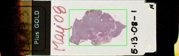
5/13/2008 Case 1: Oncocytic meningioma
Presented by: Dr. Roy Rhodes - Robert Wood Johnson University Hospital
Clinical History:
This 41 year old male had brief nocturnal new-onset seizures including changes in respiratory rate, jaw tightening, and raising the left arm. Imaging revealed a 5.3 cm left frontoparietal dural-based mass with mild edema, which was resected in 2/08 with good results and no recurrence in 3 months.
The slides showed a cellular tumor of pleomorphic cells with fibrillar cytoplasm growing as a sheet which compresses adjacent brain tissue. Nuclei contained nucleoli. There were chordoid areas and hyaluronidase removed mucoid material. The tumor expressed vimentin and EMA; KI67 labeled between 5-20% of nuclei. The anti-mitochondrial antibody stain was positive. Electron microscopy revealed desmosomes between the tumor cells and focal basal lamina formation. The choroid areas showed cells with microvillous processes in a granular proteoglycan network. Almost all (99%) of the mitochondria were abnormal with parallel flattened cristae, discoid forms, giant forms, and glycogen accumulation.
Diagnostic Notes:
The diagnosis was oncocytic meningioma. Nine cases are reported in the literature with an average age of 57.5 years. Four of the nine recurred in an average of 21 months. They are difficult to remove at surgery. Oncocytic mitochondrial changes may reflect stress, mitochondrial dysfunction or metaplasia. There is a high mutation rate in the mitochondrial DNA. Mitochondrial hypertrophy and hyperplasia may reflect and increase in manganese superoxide dismutase. The possibility that oncocytic meningioma could be a free-radical scavenging variant of chordoid meningioma was discussed.
References:
-
Harrison JD, Rose PE. Myxoid meningioma: histochemistry and electron microscopy. Acta Neuropathol 1985;68:80-82.