neuroplex
New York Association of Neuropathologists
8/12/2008 Case 2: Diffuse CNS B-cell lymphoma
Presented by: Dr. Piotr Kozlowski
Clinical History:
An 80-year-old Caucasian female become acutely ill six month prior to her demise, when she developed right hemiplegia, weakness, gait disorder, and ataxia. She was admitted to a local hospital where initial workup did not reveal a reason for the neurological abnormalities. No MRI was done since she had a pacemaker and defibrillator in place. CAT scan of the head was done but was not conclusive. Brain biopsy of the right frontal lobe was performed four months prior to death and was diagnosed as nonspecific encephalitis. A postmortem examination was requested by the decedent's family in order to establish an anatomical cause of death.
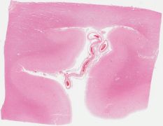
Autopsy revealed a firm granular right basal ganglia-temporal lobe lesion. A lesion in the basis pontis had the same yellow firm appearance. Microscopic examination showed a perivascular infiltrate of atypical cells spilling into the brain with occasional mitoses. This infiltrate was present in every lobe of the brain and basal ganglia, and there was a solid mass in the left cerebrum. Most of the cells were CD20-positive B-cells, though 4% were CD3-positive T-cells.
Diagnostic Notes:
The diagnosis was primary B-cell lymphoma, which represents 1-2% of all CNS tumors. Most are B-cell, non-Hodgkin type lymphoma with a predilection for the 6-7th decade of life, and increased in HIV-infected patients and those with transplants. There has been an increase in incidence in immunocompetent patients as well. While immunocompetent patients tend to have a single enhancing mass of >2 cm (70% of patients), immunodeficient patients more often have multifocal lesions (40%). Diffuse brain infiltration (as in this case) is very rare and the names lymphomatosis cerebri, microgliomatosis, reticuloendothelial sarcoma and encephalitic type lymphoma have been applied to this lesion.