neuroplex
New York Association of Neuropathologists
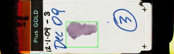
12/1/2009 Case 3: Ganglioglioma, Hemangioblastoma
Presented by: Dr. Mary Fowkes - Mount Sinai Medical Center
Clinical History:
21-year-old healthy female with one episode of loss of consciousness of unknown duration in March 2008, and with two episodes of dizziness each lasting 30 minutes in October 2008. No neurologic deficits, EEG normal. MRI showed a left frontal brain cystic mass with a mural nodule which was unchanged from 8/08 to 6/09. The cyst wall did not enhance and there was no vasogenic edema. The mural nodule showed mildly increased blood flow suggesting increased angiogenesis and/or higher grade. The radiological differential diagnosis included ganglioglioma, pleomorphic xanthoastrocytoma (PXA), metastasis, hemangioblastoma and juvenile pilocytic astrocytoma. At operation, a firm fibrotic tumor with a mural nodule was found, and frozen section was read as "consistent with glioma." As of 9/09, there was no residual neurological deficit.
Sections showed a tumor of large ganglion-like cells and a spindly component. The large cells expressed, variably, GFA, neurofilaments and synaptophysin. There were eosinophilic granular bodies and a lymphocytic perivascular infiltrate. Reticulin was present in the spindly component but not in the gangliocytic component.
Diagnostic Notes:
The diagnosis was ganglioglioma. Discussion, especially by Drs. Rosenblum and Zagzag, centered on the relationship of ganglioglioma and PXA. Are these two separate entities, or do they represent a spectrum of the same tumor entity? Both entities occur in young patients, are typically cystic, show pleomorphism with large cells and a fascicular component, express reticulin, have lymphocytes, eosinophilic granular bodies and may extend to the subarachnoid space. Neoplastic cells that look like ganglion cells (neuronal) may also express GFAP. Gangliogliomas are supposedly "white" but PXA are "yellow." The extent of resection and mitotic activity are most predictive of recurrence. Dr. Zagzag noted that the present example is a ganglioglioma because the large ganglion cells are devoid of reticulin.