neuroplex
New York Association of Neuropathologists
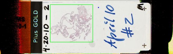
4/20/2010 Case 2: Cysticercosis (racemose form)
Presented by: Dr. Leroy Sharer - New Jersey Medical School
Clinical History:
A 49-year-old man from Guatemala, with bilateral weakness and numbness of the arms and legs. Neuroimaging revealed a multi-septated, cystic lesion in the foramen magnum region, extending into the upper cervical spinal canal, with peripheral wall enhancement but no nodules. There was mild mass effect on the medulla and the cerebellar tonsils, with apparent abnormal CSF flow at the cervicomedullary junction. There were also multiple, enhancing, calcified lesions in the cerebral hemispheres at the gray-white junctions, the largest measuring 1.6 cm in greatest dimension. The submitted material is from the posterior fossa-upper cervical cord region. A video of the removal of multiple cysts from the brainstem region was shown.
Diagnostic Notes:
The diagnosis was the racemose form of Cysticercosis, in which scolices are not seen. The calcareous bodies represent part of the organisms and are characteristic but not pathognomonic of cysticercosis. There was a focal inflammatory reaction, which occurs once the organisms die. This case had associated fibrosis and lymphoid follicles were seen. The racemose form of cysticercosis occurs in the subarachnoid space, especially around the foramen magnum.